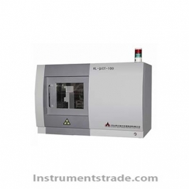
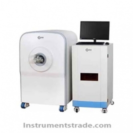
IVScope 8000X Multi Mode Small Animal Live Imaging System Introduction: The IVScope 8000X series X-ray multimodal small animal in vivo imaging system is a high-performance imaging system designed specifically for preclinical research, which can meet the needs of fluorescence, bioluminescence, and X-ray imaging. It has high sensitivity, high resolution, and X-ray radiation safety. By combining high-resolution X-ray images with high-quality optical images, researchers can obtain functional and structural imaging data to improve research design and enhance decision-making confidence, deepen understanding of disease mechanisms, disease progression, and treatment response. characteristic: 1. Multimodal imaging: bioluminescence/fluorescence/X-ray imaging 2. High sensitivity refrigerated CCD camera: The lowest temperature can reach -90 ℃ (with optional water-cooled circulation pump), which can detect extremely weak bioluminescence signals and maintain low noise 3. F/0.8 large aperture autofocus lens: ensuring sufficient light intake even in low light conditions 4. High throughput: capable of imaging up to 5 mice simultaneously 5. Powerful professional image acquisition and analysis software: used for automatic acquisition, visualization, and precise quantification of imaging data Technical parameters: Camera: 6 million pixels, front cooled CCD camera Pixel size: 4.54*4.54μm Refrigeration temperature: -30 ℃ Peak quantum efficiency: 75% Grayscale range: 16bit (0-65535) Lens: High transmittance fixed focus lens, F/0.8 large aperture, capable of automatic focusing Shooting area: 10.5cm*8.4cm ~23.5cm*18.8cm X-ray: Focus size: ≤ 0.3mm resolution: 0.2mm Overall dimensions: 620*720*1750mm(W*D*H)
Introduction: The IVScope 8000X series X-ray multimodal small animal in vivo imaging system is a high-performance imaging system designed specifically for preclinical research, which can meet the needs of fluorescence, bioluminescence, and X-ray imaging. It has high sensitivity, high resolution, and X-ray radiation safety. By combining high-resolution X-ray images with high-quality optical images, researchers can obtain functional and structural imaging data to improve research design and enhance decision-making confidence, deepen understanding of disease mechanisms, disease progression, and treatment response.