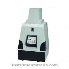
Introduction: ZF-388 automatic gel imaging analysis system adopts high-tech means of system hardware configuration: high-resolution, high-quality, high-definition CCD camera, fully automatic computer control, high degree of programmability, and digital camera can be connected with the computer Photographic imaging control, analysis software can achieve image editing processing, automatic lane identification, molecular weight calculation, sample volume distribution calculation. Guaranteed recording of DNA/RNA gels, protein gels, blotting hybridization membranes (including Western, Southern, Northern, Slot/dot blots), autoradiographic plates, microplate readers, thin-layer chromatography plates, chemical fluorescence imaging, etc. The sensitivity of the image under low illumination, without losing the band, can ultimately give the peak, molecular weight or base pair number, area, height, location, volume or total sample volume of the gel band. The greatest degree of control of EB pollution effectively protects the health of experimental operators. It helps researchers to obtain electrophoretic photographs and analysis results safely, correctly and quickly, free from tedious operations and improve work efficiency. Fully automated gel imaging system ZF-388 can be used for DNA/RNA gels, protein gels, blotting hybridization membranes (including Western, Southern, Northern, Solt, spot hybrid membranes), autoradiography, microtiter plates, thin The imaging and analysis of the layer chromatogram and other images can accurately analyze total bands, spots, and any other target area, molecular weight analysis, cluster analysis, and homology analysis. Application range: Nucleic Acid Detection: Various fluorescent dyes such as Ethidium bromide, SYBRTM Gold, SYBRTM Green, SYBRTM Safe, GelStarTM, Fluorescein, Texas Red labeled DNA/RNA detection. Protein detection: Coomassie brilliant blue glue, silver burning glue, and fluorescent dyes such as SyproTM Red, SyproTM Orange, Pro-Q Diamond, DeepPurple marker glue/membrane/chip, etc. Other applications: various hybrid membranes, protein transfer membranes, colony counts in culture dishes, microtiter plates, TLC plates, etc. Features: 1. Integrated design, built-in computer, 10.4 inch LCD computer touch screen 2. Built-in 10.4-inch computer touch screen, direct real-time observation, processing and preservation of the gel image; 3. Through the chassis panel for automatic control of transmission UV lamp and reflector lamp, manually adjust the zoom, focus, aperture, etc.; 4. Scientific-grade CCD High-definition, high-quality images embody more detail in the image and have a stronger ability to analyze weak bands. 5. Through the software or UV observation window, real-time observation of the sample, more intuitive and more secure; 6. Timing protection function: No command is input within 10 minutes, all light sources are automatically turned off, extending the service life 7. Close the operating software: The system automatically turns off all light sources at the same time, making the experiment safer and more efficient. 8. The closed chamber design provides optimal conditions for chemiluminescence and bioluminescence 9. Unique design of double-sided tapping windows on both sides: The user cuts the glue directly in the dark box to effectively prevent EB contamination. 10. Drawer light box double air cooling closed design: pull the drawer that automatically cut off the UV light source to avoid UV damage 11. Image function: adjust the image size, brightness, grayscale, contrast, angle, stripe calibration, anti-color, crop, rotate, zoom, add text 12. Integration of analysis software and image acquisition software: Image capture, analysis of electrophoresis gels, dot blots, slot blots, and colony counts were performed at the same interface. 13. Band level and verticality adjustable: The level and verticality of the band can be manually adjusted to obtain more accurate data. Technical Parameters: Patent number: 2008 3 0130298.3 Photosensitive chip: Microvision chip Camera: US Microvision high-resolution low-light digital camera Detection signal to noise ratio ≥56dB Effective pixels: 2592 (H) × 1944 (V) Resolution: 5 million pixels Acquisition bit: 16bit Lens: High-permeability electric lens, Computar 8 ~ 48mm Aperture: F1: 1.2 Automatic Exposure time: 1ms-2s Dynamic range: >4.5 orders of magnitude Detection sensitivity: Less than 20 Pg double-stranded DNA stained with EB Lighting Mode: Transmittance UV, Transmitting White, Reflecting White (Optical Blue optional) Excitation light source Transmission: 300nm, white light (optional transmission blue 200 x 120mm) Bilateral reflective white light (cold light) (R type: 254nm, 365nm UV reflection 200x50mm) Filter: Standard 590nm, Compatible with most fluorescent dyes such as EB, Sybr, GoldView Shooting area UV: 200×250mm White light: 210×260mm Tapping observation device: observation window 60×125mm, tapping window 100×125mm
Introduction: ZF-388 automatic gel imaging analysis system adopts high-tech means of system hardware configuration: high-resolution, high-quality, high-definition CCD camera, fully automatic computer control, high degree of programmability, and digital camera can be connected with the computer Photographic imaging control, analysis software can achieve image editing processing, automatic lane identification, molecular weight calculation, sample volume distribution calculation. Guaranteed recording of DNA/RNA gels, protein gels, blotting hybridization membranes (including Western, Southern, Northern, Slot/dot blots), autoradiographic plates, microplate readers, thin-layer chromatography plates, chemical fluorescence imaging, etc. The sensitivity of the image under low illumination, without losing the band, can ultimately give the peak, molecular weight or base pair number, area, height, location, volume or total sample volume of the gel band. The greatest degree of control of EB pollution effectively protects the health of experimental operators. It helps researchers to obtain electrophoretic photographs and analysis results safely, correctly and quickly, free from tedious operations and improve work efficiency. Fully automated gel imaging system ZF-388 can be used for DNA/RNA gels, protein gels, blotting hybridization membranes (including Western, Southern, Northern, Solt, spot hybrid membranes), autoradiography, microtiter plates, thin The imaging and analysis of the layer chromatogram and other images can accurately analyze total bands, spots, and any other target area, molecular weight analysis, cluster analysis, and homology analysis. Application range: Nucleic Acid Detection: Various fluorescent dyes such as Ethidium bromide, SYBRTM Gold, SYBRTM Green, SYBRTM Safe, GelStarTM, Fluorescein, Texas Red labeled DNA/RNA detection. Protein detection: Coomassie brilliant blue glue, silver burning glue, and fluorescent dyes such as SyproTM Red, SyproTM Orange, Pro-Q Diamond, DeepPurple marker glue/membrane/chip, etc. Other applications: various hybrid membranes, protein transfer membranes, colony counts in culture dishes, microtiter plates, TLC plates, etc.