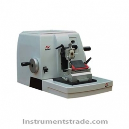
Introduction: A certain size of diseased tissue was taken and pathological sections were made by histopathological methods (usually the lesions were embedded in paraffin blocks, sliced with a slicer, and stained with hematoxylin-eosin (HE)). The microscope further examines the lesion. The pathogenesis and development of lesions, and finally make a pathological diagnosis. Features: 1. The original parts imported, the performance of the imported machine, the service of the brand machine, and the higher cost performance. 2. Using the international mainstream mature technology, innovative quality design, complete elimination of jumping knife, uneven thickness, and the phenomenon of inconsistent knife and knife, establish a new image of the Chinese slicer brand. 3. The R135 adopts the mature technology of the mainstream slicer and can obtain the best slice effect. It is the first choice for workers who are accustomed to using steel knife slicing. 4. The streamlined cover does not need to be disassembled, so it is easy to clean; the top tray can be used to place items such as specimen boxes. 5. The sample holding system can be arbitrarily adjusted on the three-dimensional axis and is easy to use. 6. Eliminating completely the phenomenon of jumping knife, thick book, uneven knife, knife, etc. 7. Whether you use a standard slicing knife or disposable blade, you can get the ideal slice effect. 8. Compatible with most of the accessories imported mainstream slicing machine, providing guarantee for value upgrade.
Introduction: A certain size of diseased tissue was taken and pathological sections were made by histopathological methods (usually the lesions were embedded in paraffin blocks, sliced with a slicer, and stained with hematoxylin-eosin (HE)). The microscope further examines the lesion. The pathogenesis and development of lesions, and finally make a pathological diagnosis.